Biotinylated Human Chemokine Proteins
The versatility and specificity of biotin-streptavidin chemistry make biotinylated chemokines valuable tools in studying chemokine biology and related research areas. Utilize our products to design and develop custom assays in areas of cancer biology, immunology, and infectious diseases. Here are some examples of common applications with our biotinylated chemokines:
Receptor Binding & Internalization Studies • Cell Migration Assays • Affinity Purification • Imaging and Visualization • Drug Discovery • In-Vivo studies
Our recombinant bacterial system has been optimized to ensure endogeneous functional activity. We diligently test our products to ensure their activity with low endotoxin levels (<0.01 EU per 1μg). For more information, click on the products below for detailed specifications.
Biotinylated Chemokine Products
Application Data
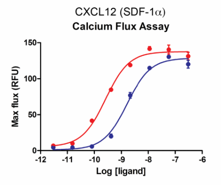
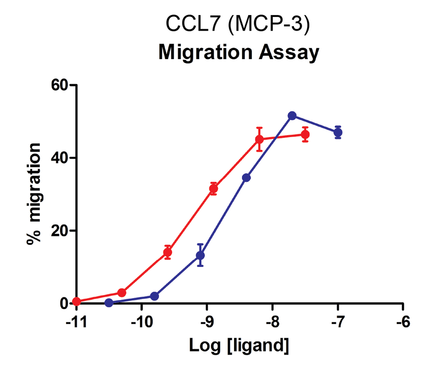
Activity Assays - Calcium Flux and Cell Migration
|
U937 cells expressing endogenous CXCR4 were stimulated with increasing concentrations of SDF-1α after a 90-minute incubation with calcium dye at 37oC. The maximum response was plotted as a function of the ligand concentration (Red: wild type; Blue: biotinylated).
|
Cells expressing recombinant CCR2 were assayed for migration through a transwell bare filter at various concentrations of MCP-3. The responses are expressed as the % of total input cells (Red: wild type; Blue: biotinylated).
|
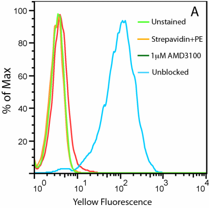
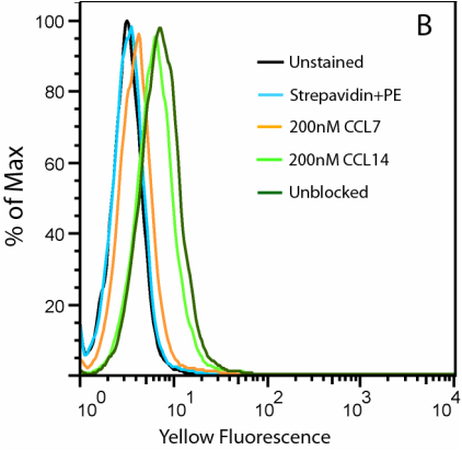
Cell Surface Expression with Flow Cytometry
|
A. Uptake of CXCL12-biotin by U937 cells in the presence (red trace) and absence (cyan) of a CXCR4 inhibitor, AMD 3100. U937 cells are not stained by streptavidin-PE in the absence of CXCL12-biotin (orange).
|
B. Uptake of CCL2-biotin by THP1 cells (dark green trace) is abolished by the addition of CCL7 (orange) but not CCL14 (light green), suggesting a CCR2-specific internalization. THP1 cells are not stained in the absence of CCL2-biotin (cyan).
|
Imaging Applications - Fluorescent Cell Surface Detection
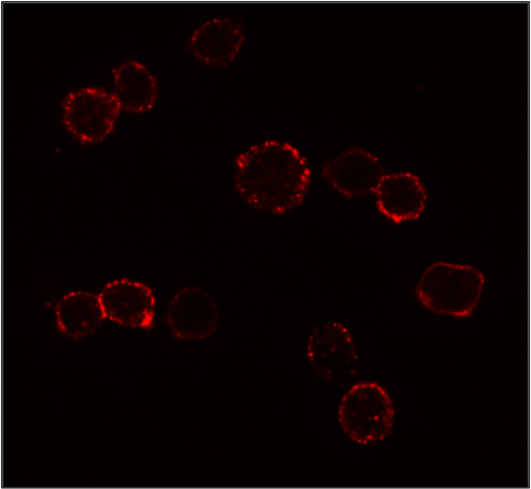
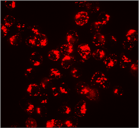
Receptor Internalization Studies - Temperature Dependent CXCR4
Distribution of the endogenous CXCR4 in Jurkat cells at 4°C (left image) and their internalization upon stimulation at 37°C (right image) were revealed by the biotinylated SDF-1α bound by streptavidin-Cy3 (red).
|
Want to learn more about assays with biotinylated Chemokines? Check out our new protocols |
Biotinylated Chemokine Products

|

Human CXCL12 (SDF-1α) Biotinylated
Lyophilized sample, >97% Purity, EC50 = 2.5nM |
|
Purchase directly on product pages or request a quote for more information.
|

