Recombinant Biotinylated Chemokine Proteins from ChemoTactics
  • Chemokine Products
    • Biotinylated Chemokines >
      • Biotinylated CCL2 (MCP-1)
      • Biotinylated CCL3 (MIP-1α)
      • Biotinylated CCL4 (MIP-1β)
      • Biotinylated CCL5 (Rantes)
      • Biotinylated CCL7 (MCP-3)
      • Biotinylated CCL14 (HCC-1)
      • Biotinylated CCL19 (MIP-3β)
      • Biotinylated CCL28 (MEC)
      • Biotinylated CXCL8 (IL-8)
      • Biotinylated CXCL12 (SDF-1α)
    • Unmodified Chemokines >
      • CCL2 (MCP-1)
      • CCL3 (MIP-1α)
      • CCL4 (MIP-1β)
      • CCL5 (RANTES)
      • CCL7 (MCP-3)
      • CCL14 (HCC-1)
      • CCL19 (MIP-3β)
      • CCL27 (CTACK)
      • CCL28 (MEC)
      • CXCL8 (IL-8)
      • CXCL10 (IP-10)
      • CXCL12 (SDF-1α)
  • Streptavidin Conjugates
  • Services
  • Resources
    • Published Applications Biotinylated CXCL12 and CCL5
    • What are Chemokines?
    • Chemokine Protocols >
      • Flow Cytometry with Biotinylated Chemokines
      • ELISA with Biotinylated Chemokines
      • Transwell Migration Assay
    • Blogs
  • About
    • Contact

Biotinylated Human Chemokines

Picture
The versatility and specificity of biotin-streptavidin chemistry make biotinylated chemokines valuable tools in studying chemokine biology and related research areas. ​Utilize our products to design and develop custom assays in areas of cancer biology, immunology, and infectious diseases. Here are some examples of common applications  with our biotinylated chemokines: 

Applications

• Flow Cytometry 
• Protein-Protein Interactions
• Receptor Binding & Internalization Studies
• Cell Migration Assays
• Affinity Purification
• Surface Plasmon Resonance (SPR)
• Nanoparticle conjugation
• Imaging and Visualization
• Drug Discovery
​• In-Vivo studies
​Our recombinant bacterial system has been optimized to ensure endogeneous functional activity. We diligently test our products to ensure their activity with low endotoxin levels (<0.01 EU per 1μg). For more information, click on the products below for detailed specifications. 

Biotinylated Human Chemokine Proteins

B-CCL2
Biotin-CCL2 (MCP-1)
B-CCL2
B-CCL3
Biotin-CCL3 (MIP-1a)
B-CCL3
B-CCL4
Biotin-CCL4 (MIP-1B)
B-CCL4
B-CCL5
Biotin-CCL5 (Rantes)
B-CCL5
B-CCL7
Biotin-CCL7 (MCP-3)
B-CCL7
B-CCL14
Biotin-CCL14 (HCC-1)
B-CCL14
B-CCL19
Biotin-CCL19 (MIP-3B)
B-CCL19
B-CCL28
Biotin-CCL28 (MEC)
B-CCL28
B-CXCL8
Biotin-CXCL8 (IL-8)
B-CXCL8
B-CXCL12
Biotin-CXCL12 (SDF-1a)
B-CXCL12

Request a Quote

Request A Quote
Application Data

Activity Assays - Calcium Flux and Cell Migration

Picture
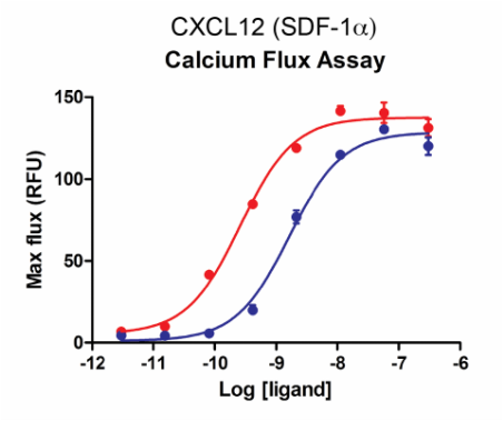
U937 cells expressing endogenous CXCR4 were stimulated with increasing concentrations of SDF-1α after a 90-minute incubation with calcium dye at 37oC.  The maximum response was plotted as a function of the ligand concentration (Red: wild type; Blue: biotinylated).  
Picture
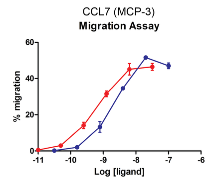
Cells expressing recombinant CCR2 were assayed for migration through a transwell bare filter at various concentrations of MCP-3.  The responses are expressed as the % of total input cells (Red: wild type; Blue: biotinylated).


Cell Surface Expression with Flow Cytometry

Picture
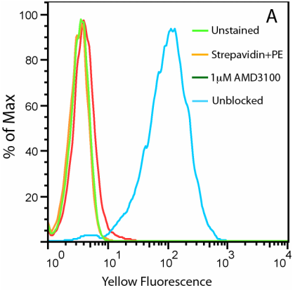
A.  Uptake of CXCL12-biotin by U937 cells in the presence (red trace) and absence (cyan) of a CXCR4 inhibitor, AMD 3100.  U937 cells are not stained by streptavidin-PE in the absence of CXCL12-biotin (orange).

Picture
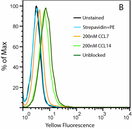
B.  Uptake of CCL2-biotin by THP1 cells (dark green trace) is abolished by the addition of CCL7 (orange) but not CCL14 (light green), suggesting a CCR2-specific internalization.  THP1 cells are not stained in the absence of CCL2-biotin (cyan).    

Imaging Applications - Fluorescent Cell Surface Detection 

Visualization of GAG Binding 
Picture

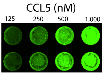
Detection of chemokine GAG binding on the cell surface using increasing amounts of biotinylated CCL5. 
Western Detection of IL-8
Picture
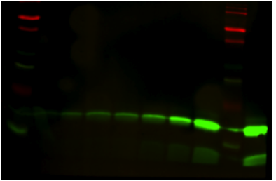
Detection of various amounts of biotinylated CXCL8 (IL-8) using streptavidin conjugated to 800W dye. 

Receptor Internalization Studies - Temperature Dependent CXCR4 

Distribution of the endogenous CXCR4 in Jurkat cells at 4°C (left image) and their internalization upon stimulation at 37°C (right image) were revealed by the biotinylated SDF-1α bound by streptavidin-Cy3 (red).
4°C
Picture
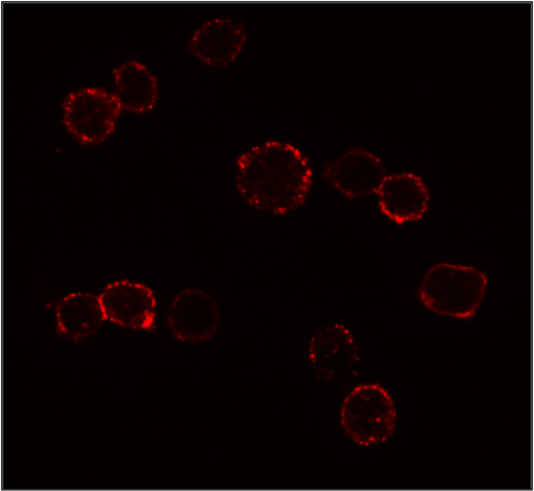
In presence of biotinylated SDF-1α bound by streptavidin-Cy3 (red), the majority of the CXCR4 population is located at the plasma membrane at 4°C. 
37°C
Picture
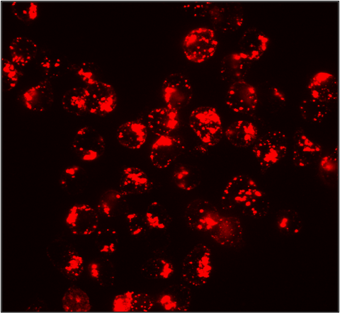
The majority of the population of CXCR4 at 37°C in the presence of biotinylated SDF-1α bound by streptavidin-Cy3 (red) has been internalized. 

Streptavidin Fluorescent Conjugates

Streptavidin - DyLight 405

Pricing

Streptavidin - DyLight 488

Pricing

Streptavidin - DyLight 550

Pricing

Streptavidin - DyLight 594

Pricing

Streptavidin - DyLight 650

Pricing
Create flexible assays by pairing biotinylated chemokines with fluorescent streptavidin conjugates. Depending on your applications and instruments different colors are more advantageous. Conveniently change out your colors with a single biotinylated chemokine. See our list of Streptavidin conjugates here. 
Streptavidin Conjugates

Want to learn more about assays with biotinylated Chemokines?
Check out our new protocols   
Chemokine Protocols
Purchase directly on product pages or request a quote for more information. 
Request A Quote

Publications

  1. Pisani, A., Donno, R., Valenti, G., Pompa, P. P., Tirelli, N., & Bardi, G. (2022). Chemokine-decorated nanoparticles target specific subpopulations of primary blood mononuclear leukocytes. Nanomaterials, 12(20), 3560. https://doi.org/10.3390/nano12203560
  2. Pisani, A.; Donno, R.; Gennari, A.; Cibecchini, G.; Catalano, F.; Marotta, R.; Pompa, P.P.; Tirelli, N.; Bardi, G. CXCL12-PLGA/Pluronic Nanoparticle Internalization Abrogates CXCR4-Mediated Cell Migration. Nanomaterials 2020, 10, 2304. https://doi.org/10.3390/nano10112304
  3. Wang Z, Moresco P, Yan R, Li J, Gao Y, Biasci D, Yao M, Pearson J, Hechtman JF, Janowitz T, Zaidi R, Weiss MJ, Fearon DT. Carcinomas assemble a filamentous CXCL12-keratin-19 coating that suppresses T cell-mediated immune attack.  PNAS Jan 2022 Vol. 119 No. 4.  https://doi.org/10.1073/pnas.2119463119
  4. Chatterjee, T., Johnson-Buck, A. Walter, NG. Highly sensitive protein detection by aptamer-based single-molecule kinetic fingerprinting. Biosensors and Bioelectronics. Vol 216 15 Nov 2022. https://doi.org/10.1016/j.bios.2022.114639
  5. Zhikai Wang, Ran Yan, Jiayun Li, Ya Gao, Philip Moresco, Min Yao, Jaclyn F. Hechtman, Matthew J. Weiss, Tobias Janowitz, Douglas T. Fearon. Pancreatic cancer cells assemble a CXCL12-keratin 19 coating to resist immunotherapy. bioRxiv 776419; doi: https://doi.org/10.1101/776419. Posted: September 4, 2020
  6. Torretta S., Colombo G., Travelli C, Boumya S., Lim D., Genazzani A., Brolla A. The Cytokine Nicotinamide Phosphoribosyltransferase (eNAMPT; PBEF; Visfatin) Acts as a Natural Antagonist of C-C Chemokine Receptor Type 5 (CCR5). Cells 2020 Feb; 9(2):496. doi 10.3390/cells9020496 PMID: 32098202
  7. Wu, B., Chien, E.Y., Mol, C.D., Fenalti, G., Liu, W., Katritch, V., Abagyan, R., Brooun, A., Wells, P., Bi, F.C., Hamel, D.J., Kuhn, P., Handel, T.M., Cherezov, V., Stevens, R.C. Structures of the CXCR4 chemokine GPCR with small-molecule and cyclic peptide antagonists.  Science, 2010. 330: 1066-1071. PMID: 20929726
  8. Ervin E. Kara, Iain Comerford, Cameron R. Bastow, Kevin A. Fenix, Wendel Litchfield, Tracey M. Handel, and Shaun R. McColl.  Distinct chemokine receptor axes regulate T helper 9 cell trafficking to allergic and autoimmune inflammatory sites. J Immunol., 191:1110-1117, 2013. PMID: 23797668
  9. M. O'Hayre, C.L. Salanga, T.J. Kipps, D. Messmer, P.C. Dorrestein, T.M. Handel (2010) Elucidating the CXCL12/CXCR4 Signaling Network in Chronic Lymphocytic Leukemia through Phosphoproteomic Analysis. PLoS One. 5:e11716. PMID: 20661426
  10. T.M. Handel, Z. Johnson, M. Mack, R. Cirillo, V. Muzio, M. Teixiera, M. Déruaz, F. Borlat, T.N.C. Wells, and A.E.I. Proudfoot (2008). An Engineered Monomer of CCL2 has Anti-inflammatory Properties Emphasizing the Importance of Oligomerization for Chemokine Activity In Vivo. J Leukocyte Biology. 84(4):1101-8. PMID: 18662971​
Picture
​ChemoTactics, Inc
6076 Corte del Cedro Carlsbad,
​CA 92011 USA
Phone: +1-(858)-412-0485
​Email: [email protected]
Chemokine Products
Biotinylated Chemokines
Unconjugated Chemokines
Streptavidin Conjugates
Research Services
Resources​
Published Applications
Protocols
​Blog
About
​
Contact
Made in the USA
  • Chemokine Products
    • Biotinylated Chemokines >
      • Biotinylated CCL2 (MCP-1)
      • Biotinylated CCL3 (MIP-1α)
      • Biotinylated CCL4 (MIP-1β)
      • Biotinylated CCL5 (Rantes)
      • Biotinylated CCL7 (MCP-3)
      • Biotinylated CCL14 (HCC-1)
      • Biotinylated CCL19 (MIP-3β)
      • Biotinylated CCL28 (MEC)
      • Biotinylated CXCL8 (IL-8)
      • Biotinylated CXCL12 (SDF-1α)
    • Unmodified Chemokines >
      • CCL2 (MCP-1)
      • CCL3 (MIP-1α)
      • CCL4 (MIP-1β)
      • CCL5 (RANTES)
      • CCL7 (MCP-3)
      • CCL14 (HCC-1)
      • CCL19 (MIP-3β)
      • CCL27 (CTACK)
      • CCL28 (MEC)
      • CXCL8 (IL-8)
      • CXCL10 (IP-10)
      • CXCL12 (SDF-1α)
  • Streptavidin Conjugates
  • Services
  • Resources
    • Published Applications Biotinylated CXCL12 and CCL5
    • What are Chemokines?
    • Chemokine Protocols >
      • Flow Cytometry with Biotinylated Chemokines
      • ELISA with Biotinylated Chemokines
      • Transwell Migration Assay
    • Blogs
  • About
    • Contact